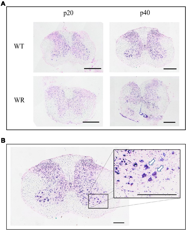
Fig. 2. Expression pattern of NDRG2 mRNA in the cervical spinal cord of wild type and wobbler mice. (A) Exemplary overview of in situ hybridization with NDRG2-probe in a cross section of the cervical spinal cord of wild type (WT) and wobbler (WR) mice at the developmental stage p20 and p40. There are no visible staining pattern differences between WT and WR. Scale bar = 1 mm. (B) Cross section of the cervical spinal cord of a wild type mouse at p40. Exemplary large-scale picture of the anterior horn after in situ hybridization with NDRG2-probe. Arrows indicate motor neurons with a clearly visible prominent staining for NDRG2. Scale bar = 500 µm. All pictures were taken with a light microscope (Olympus microscope BX61VS, Japan) and a 20x objective (UPlanSApo 20x/0.4, Olympus, Japan). For counterstaining Nuclear Fast Red was used.